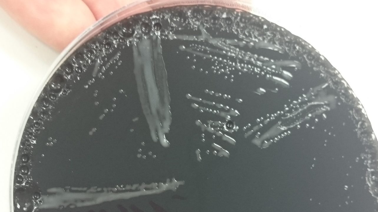
Are Coliforms and Enterobacteriaceae the same thing?

Back

Some interesting facts about Enteros and Coliforms

January 18, 2018

Express Micro Science

If you look up Coliforms and Enterobacteriaceae in Wikipedia and read the first two lines:

“Coliform bacteria are defined as rod-shaped Gram-negative non-spore forming and motile or non-motile bacteria which can ferment lactose with the production of acid and gas when incubated at 35–37°C. They are a commonly used indicator of sanitary quality of foods and water.”

“The Enterobacteriaceae are a large family of Gram-negative bacteria that includes, along with many harmless symbionts, many of the more familiar pathogens, such as SalmonellaEscherichia coliYersinia pestisKlebsiella, and Shigella. Other disease-causing bacteria in this family include ProteusEnterobacterSerratia, and Citrobacter.”

One might get the impression these are two completely different groups of bacteria. That may be how Wikipedia works, one person wrote one article and another wrote the other so the style is somewhat different. In any case, when testing food in a microbiology lab, there isn’t a whole lot of difference between the two.

Both Coliforms and Enterobacteriaceae (Enteros) are used as indicator organisms in microbiology.  Most of them do not cause illness but where they come from, the environments in which they grow well and the way they die is the same for most foodborne pathogens. Therefore it is a good indication that if these bacteria are high in a food the likelihood of more sinister bacteria being present increases and vice versa.

But can these bacteria groups really be used interchangeably?

In the laboratory we use slightly different agars to grow Coliforms and Enteros, the difference being Coliforms metabolise lactose better than glucose so we use lactose as the main sugar when testing for Coliforms (Violet Red Bile Agar) and glucose for Enteros (Violet Red Bile Glucose Agar). The theory being not all Enteros can metabolize lactose but all Coliforms can metabolize glucose so the Coliform media is slightly more selective than the Entero media.

But how does this effect your test report?  If you asked for both Coliforms and Enteros on the same sample, it would depend on the sample type if any real differences in the result would ensue.

For example, dairy products primary sugar is lactose so Coliforms grow better in these products and should probably be grown up on a lactose based agar. In meat where glucose is more prevalent but always at a low level overall, the number of Coliforms and Enteros would probably be very similar. In fact most other foods glucose or sucrose, which contains glucose, is much more common so the more general VRBGA should be used to capture as many indicator bacteria as possible.


                              Top Facts

  1. All coliforms are enteros but not all enteros are coliforms
  2. Coliforms prefer lactose as their sugar source but can metabolize glucose
  3. Not all enteros can grow on lactose alone but most can
  4. Both coliforms and enteros are good indicator organisms for sanitation in water and food, as they come from the same places, grow well under the same conditions and are killed in similar fashion as many pathogens


For Coliforms, Enteros and other microbiology testing available from Express Micro Science, please click on food testing.

More information on indicator organisms will be posted soon…

Salmonella enteritidis and testing for the vaccine strain

Back

Why PCR testing helps with Salmonella enteritidis differentiation

January 5, 2018

Salmonella enterica subsp. enterica is a rod-shaped, facultatively anaerobic gram negative bacterium that has several subtypes called serovars, which are the primary cause of Salmonella infections in humans, also known as salmonellosis. Salmonellosis is the second most common foodborne disease after campylobacteriosis. Common symptoms are diarrhoea, fever, vomiting and abdominal cramps 12 to 72 hours after infection.

One serovar, Enteritidis, is responsible for over 40% of total recorded human infections worldwide. The primary reason is the vertical transmission or zoonosis of Salmonella enteritidis between galliformes (ground-feeding birds, such as chickens, turkeys and pheasants) and humans because of the worldwide consumption of poultry meat and associated products e.g. eggs.

Between 1981 and 1991 the numbers of Salmonella infections rose by 170% in the UK, primarily due to an epidemic of Salmonella enteritidis. An industry-led vaccination scheme began in breeding flocks in 1994 and in laying flocks in 1998. Mass vaccination has continued for breeders subscribing to the Lion Quality Code of Practice and using the Lion Mark on eggs, which account for approximately 85% of the UK market. As a result of this and other control measures, the number of cases fell eight-fold from 1.6 cases per 1000 persons per year from 1993 to 1996 to just 0.2 cases per 1000 persons per year in 2008 to 2009. The number of lab-confirmed cases dropped from 18000 in 1993 to just 459 in 2010.

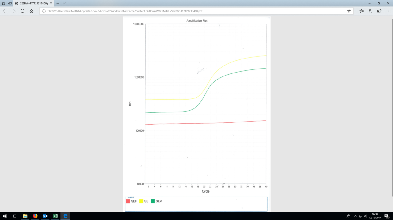
There are several vaccine strains of commercial Salmonella enteritidis available on the market. However, in traditional culture techniques for detection of Salmonella, they cannot differentiate between these harmless vaccine strains and the harmful ‘field’ strains of Salmonella enteritidis that may be present. In order to differentiate the two, we have to use the technique polymerase chain reaction (PCR). PCR amplifies a copy of a small segment of DNA exponentially to generate millions of copies of a particular DNA sequence. This DNA sequence is unique to the vaccine strains of Salmonella enteritidis and the detection of it can then be used to differentiate from the field strains.


Express Micro Science utilises two different PCR kits that can detect and differentiate different commercial vaccine strains of Salmonella enteritidis such as Salmovac SE, Salmovac 440 Gallivac SE and AviPro SALMONELLA VAC E. For more information on the testing we can perform on poultry, please contact us.


Written by: Paul Moffat, Microbiologist and Project Manager at Express Micro Science


Christmas Bugs – Clostridium perfringens

Back

What to do to avoid getting Clostridium perfringens this Christmas

December 22, 2017

Christmas is for eating! Eating large meals sharing with lots of people. These meals are not our usual day to day fare and if you’re not careful could lead to the case of a bad tummy bug, Clostridium perfringens – which could ruin all the holiday cheer for days or even weeks.

So, what to watch out for when preparing your feasts for your families this year?

Clostridium perfringens

A little-known bacterium called Clostridium perfringens can raise its scary head at Christmas time. Clostridium perfringens is a Gram-positive, rod-shaped, anaerobic, spore-forming pathogenic bacterium [1] . C. perfringens is the third most common cause of food poisoning in the United Kingdom though it can sometimes be ingested and cause no harm [2].

So how does it give us problems at Christmas time? Well it happily lives in the intestines of poultry (e.g. Turkey), which is a nice anaerobic (oxygen-free) environment and so comes into the house inside the large turkey purchased for Christmas dinner. C. perfringens is a member of the Clostridium species it is also a spore former which means it can withstand very high temperatures and so will be killed during cooking of the turkey – (even well-cooked turkeys so don’t blame the chef on this one!)

How it gets us, is how we handle the turkey after Christmas dinner is over…..

Here’s a scenario, everyone is full, and no one can be bothered cleaning up; let alone cut up the turkey and store the leftovers safely in the refrigerator. So instead the remains of the full carcass are left on the counter – cooling slowly in the lovely warm kitchen. No one even thinks about that turkey until late Christmas night when a nice cold turkey sandwich sounds appetising and this is where the danger lies. Clostridium perfringens multiply extremely fast in just 6.5 minutes under optimum conditions, within the short period of time of Christmas lunch to late night snack their levels can get dangerously high [3]. With no reheating one can ingest the toxin produced by these bacterium and symptoms typically abdominal cramping, diarrhoea, vomiting, and fever will ensue. The whole course usually resolves within 24 hours, but can last up to two weeks or even death in older or infirm people.

So how to avoid this disaster? Several things could have been done better.

  1. Cook your turkey following the instructions and make sure its internal temperature reaches 75C or above – eat whilst it’s still hot!
  2. Shortly after finishing the meal, make sure the turkey, stew or other large dish of leftovers is subdivided into smaller portions and stored in the refrigerator (Perfringens species do not do well at cold temperatures and won’t grow at all in the presence of oxygen)
  3. Reheat food which may have been subjected to slow cooling to piping hot – this will at least deactivate the toxin that C perfringens produce which is the actual culprit for making us sick

Follow those simple three rules and you and your family will avoid food poisoning from C perfringens, but beware there are two other pathogens closely linked to poultry – Salmonella and Campylobacter – for more information on those bad guys see our blogs ……

Examples of Clostridium perfringens outbreaks

On May 7, 2010, “42 residents and 12 staff members at a Louisiana state psychiatric hospital were affected experienced vomiting, abdominal cramps, and diarrhea”. Three patients died within 24 hours. The outbreak was linked to chicken which was cooked a day prior to being served and was not cooled down according to hospital guidelines. The outbreak affected 31% of the residents of the hospital and 69% of the staff who ate the chicken. It is unknown how many of the affected residents ate the chicken. [5]
In January 2017, a restaurant in, New York was sued when 260 people were sickened after eating foods contaminated with Clostridium perfringens. “Officials from the Monroe County Department of Public Health closed down the Golden Ponds after more than a fourth of its Thanksgiving Day guests became ill. An inspection revealed a walk-in refrigerator with food spills and mould, a damaged gasket preventing the door from closing, and mildew growing inside.[6]:


References

  1. https://en.wikipedia.org/wiki/Clostridium_perfringens
  2. Ryan, Kenneth J.; Ray, C. George (2004). Sherris Medical Microbiology: an Introduction to Infectious Diseases (4th ed.). New York: McGraw-Hill. p. 310. ISBN 0-8385-8529-9.
  3. The Essentials of Food Microbiology, p31, John Garbutt, pub Hodder Arnold 1997
  4. “Fatal Foodborne Clostridium perfringens Illness at a State Psychiatric Hospital — Louisiana, 2010″.Centers for Disease Control and Prevention. Retrieved 16 November 2013.
  5. “Mother, son sue eatery for Thanksgiving dinner food poisoning – Food Safety News”. 6 January 2017


Express Micro Science conducts a range of microbiology food testing to help manufacturers identify bacteria in foods. We can test for a range of pathogenic and indicator organisms to look at specific bacteria and to help monitor factories. For more information please click on food testing to see the testing available from us, or please contact us directly.


Picture courtesy of: https://www.express.co.uk/life-style/health/889395/food-poisoning-symptoms-christmas-cook-turkey-bacteria

Worms in Chickens and Why Worm Counts are Important

Back

Worms and worm counts explained

December 18, 2017

This article explains about worms in chickens and why performing worm counts is useful for farmers to monitor the health of their chickens.

We regularly worm our cats, dogs, horses and cows; why not worm our chickens?

Worms in chickens can be divided into two major groups. The first group most commonly affect the gastrointestinal tract of chicken. The second group is gape worms which live in a bird’s trachea.

There are 4 species of worms which are found in gastrointestinal tract of bird’s:

  • Capillaria
  • Ascaridia
  • Heterakis
  • Tapeworms

Capillaria (Threadworms)

This is the smallest of all and most dangerous if present in high numbers. They grow up to 1.5cm and found mostly in lower intestinal tract. Capillaria causes diarrhea, anaemia, inflammation, hemorrhage and bird’s look hunched and dull. This results in decreased egg production and low fertility. In severe cases it leads to death.

Ascaridia

This is the most common and largest worm in chickens which can grow up to 7 cm and lives in the small intestine of chickens. These worms cause severe damage to gut lining and result in reduced food uptake. Highly infected birds may cause diarrhoea  lethargy, and reduced egg production.

Heterakis

This worm is also called as caecal worm as it is most commonly found in caecum of chickens. Presence of this worm is non pathological but chickens can act as vectors for blackhead disease.

Tapeworms

Tapeworms rarely affect chickens.

Gapeworms

Gapeworms are commonly called as Syngamus trachea, they are roundworms and live in trachea of chickens. Affected birds have open mouthed breathing and results in difficulty in breathing. In severe cases it causes suffocation and leads to death.

Oocysts

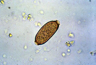
When we observe bird droppings there will be a presence of coccidiosis. Coccidiosis is a parasite which damages the gut walls of chickens. There are six different species of coccidiosis. Each of these species may be working together to cause disease.

The Oocyst has a very thick wall and it is resistant from heat, cold and disinfectants. When eaten by the chicken, it is broken down by chemicals in the gut and releases the infective form of coccidiosis. Eventually this results in production of more oocysts and passed out in faeces and infect other chickens.

Mites

Chickens are very susceptible to mite infestation. Mites can crawl up to human and can cause irritation.

What can worms do to birds?

They can cause a variety of problems:

  • Loss of shell colour and strength, yolk colour and eg size.
  • Poor appetite
  • Poor body weight, dull and pale combs
  • Risk of egg peritonitis
  • Death in high infestations

Therefore performing a worm count is important for farmers as it helps them monitor the health of the birds.

So what is a worm count?

  • A worm count is an examination of faeces under a microscope. A count is taken of the number of worm eggs present and the results are used to monitor the worm burden.
  • The results are represented as eggs per gram of faeces.
  • The number of eggs is a representation of adult worms in the gut of chicken.

Express Micro Science routinely carries out worm count testing so that poultry farmers can control their birds health through worming treatment. This keeps their birds healthy and their egg production high. We also offer Salmonella testing for poultry and eggs to meet the British Lion Code of Quality.  These tests are of course, UKAS accredited and we have been approved by both the Department for Environment, Food, and Rural Affairs (DEFRA), and the Scottish Government.


Written by: Saichalam Puttagunta, Microbiologist at Express Micro Science


Christmas and New Year Arrangements

Back

We will be open for testing

December 8, 2017

As the festive season fast approaches, we wanted to make sure you are all aware of our operating provisions during the Christmas and New Year period.

  • Our lab will be open each day over Christmas and New Year
  • Collections will not be available on 25th and 26th December or 1st and 2nd January – normal pick ups are available as usual for the other days
  • The offices will be closed on 25th and 26th December and 1st and 2nd January

If you require testing over the festive period, please contact us to let us know your requirements. This will ensure the lab is ready for your samples.


Lastly and definitely not least – wishing everyone peace, goodwill and happiness – the basic ingredients for a Happy Christmas!!


From all the staff at Express Micro Science

3 Quick Checks to Help you Understand your Water Results

Back

Your water testing results explained

November 30, 2017

With water testing being an important part of any testing programme, I thought it would be useful to include some guidance to help you understand your water reports. Below are therefore some quick checks you can do.

The standard 4 tests for water testing are TVCs at two temperatures 22OC and 37OC, Coliforms and E. coli. Sometimes you might test for Enterococci (sometimes called Faecal Streptococcus) and/or Sulphite Reducing Clostridium (or Clostridium perfringens if you like). And other water samples like pools, spas, cooling towers etc will require Pseudomonad and Legionella testing.

No. 1 – Why Test at 22OC and 37OC?
TVCs (Total Viable Count, also known as ACC or APC – please see previous blog post – TVC, ACC, APC what’s the difference?) is the number of bacteria we can get to multiply under aerobic (with oxygen) conditions, at 22OC within 72 hours or 37OC within 48 hours. The major difference between these tests is the temperature that the plates are incubated. The theory is the 22OC count will be more indicative of the resident microflora whilst the water is in the pipe, bottle etc – as this is considered “room temperature”. The 37OC will tell you if there are bacteria in the water which prefer to grow at body temperature and thus may be more sinister if the water is drunk. Very often if the TVCs are high both tests show high counts, but if only one is high it is generally the 22OC that is higher than the 37OC – not a hard and fast rule but a good indicator of reliability; see too much of this and start asking questions.

No. 2 – All E. coli are Coliforms
All E. coli are Coliforms so it is not possible to have E. coli with no Coliforms – but it is possible (and pretty common) to have Coliforms and no E. coli. And unlike when testing food for E. coli and Coliforms it is the same test for both results, so it really is impossible to have more E. coli than Coliforms in a water sample.

No. 3 – Coliforms but no TVCs…how is this possible?
We get asked how can we have Coliforms (or E. coli, or Enterococci or SRBs) in our water when there are no TVCs? This can happen and actually happens on a fairly regular basis. The reason is these tests are much more sensitive than the TVC test. The TVC test only uses 1ml of water where these other tests use 100ml of water – They are 100 times more sensitive!


Hope this helps you better understand your water reports. If you have any questions or comments please send them to me: jennifer.newton@expressmicroscience.co.uk


Express Micro Science can perform a range of microbiological tests for your water samples. Please click on EMS water testing to see the complete range of tests available, including Legionella testing and testing potable water and spas.


3 Quick checks to ensure believable micro results

Back

How do you know if your micro results make sense?

November 20, 2017

Sometimes the amount of data your microbiology lab sends you ends up hiding the information. So many not detected or “<” (less thans) it can be hard to be sure all is correct. We also know those pesky bacteria don’t always follow hard and fast rules like chemical compounds are more likely to do – so when do you feel justified to challenge your lab on their micro results?

Here are a few red flags to watch out for:

Quick Check #1
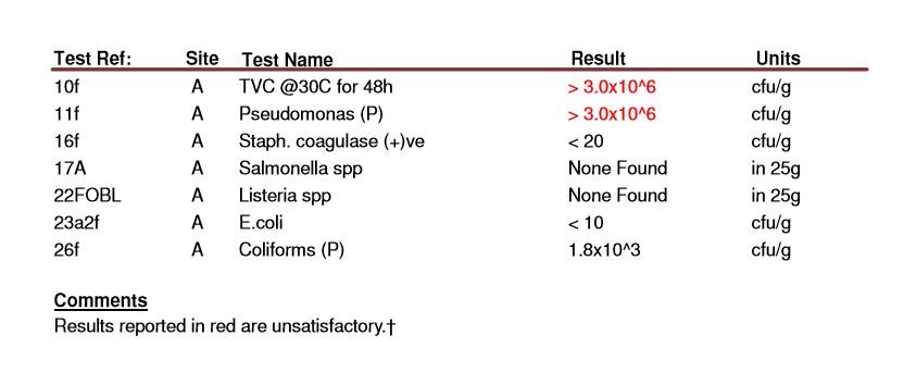
TVC result should be higher than your Coliforms, Enterobacteriaceas (Enteros) and E coli results

TVC (total viable count, also known as ACC or APC (see a previous blog post) is the number of bacteria we can get to multiply under aerobic (with oxygen) conditions, at 30C within 48 (or 72) hours. There may be other bacteria there which require other growth conditions but this is the method for TVC counts in food. So if in theory all the aerobic bacteria should be detected on the TVC plate, therefore you should not find Enterobacteriaceae, Coliforms, Staphylococcus species, Pseudomonads, E coli or other aerobes (bacteria capable of growing in the presence of oxygen) counts being higher than your TVC result.

Quick Check #2
Coliform and Enterobacteriaceae results should be pretty much the same and both should have higher results than E coli

All Coliforms are Enteros (Enterobacteriaceae) but not all Enteros are Coliforms. Most people don’t test for both at the same time because they are almost interchangeable; but should you get a significantly higher result for Coliforms over Enteros something is not right (I would accept up to 100 Coliforms to < 10 Enteros, but not 1000 Coliforms and 100 Enteros!). Another thing – E coli are Coliforms and thus also part of the Enterobacteriaceae group; and since they are just one species within this very large group it is very rare for a lab to find E coli and no Coliforms or Enteros! *


Quick Check #3
Where there are a lot of Lactics and Pseudomonads there should be a lot of TVCs 3.

This is a little bit harder to explain and not as black and white as Quick Checks 1 and 2, but still one to watch out for. Since Lactic acid bacteria can grow aerobically and Pseudomonads are strict aerobes they are capable of growing on a TVC test (see quick check fact #1). So if there are a lot of lactics – you should have a lot of TVC and the same for Pseudomonads. The only thing is there may be lots of other aerobic bacteria so the TVC result could be much higher and because the agar used to grow the Lactics and Pseudomonads is specially prepared for these bacteria they can sometimes be better at recovering these bacteria than the non-speciifc agar used in the TVC test. But again if you see a > 1000 Pseudomonads and no TVC….. It may be worth requesting a retest.

Now for a few things you cannot assume:

1. You cannot assume that if there are no Enterobacteriaceae there will not be any Salmonella, even though Salmonella are also Enteros. The tests for Salmonella are much more sensitive than that for Enteros – so although the chance that Salmonella is present goes up the more Enteros you have, having none doesn’t rule the risk out.
2. Having no E coli means there is no E coli O157. E coli are indicator organisms which won’t make anyone sick but E coli O157 is one strain which does make people very sick. Testing only for E coli cannot rule out the absence of E coli O157 for two reasons: (1) the same as in the case of Salmonella testing for E coli O157 is much more sensitive than testing for E coli in general. (2) E coli O157 actually isn’t detectable on the routine E coli test! For more information on this watch out for our blog on E coli O157 coming soon.

And one final word of warning: Microbiology deals with living things so they don’t always follow hard and fast rules, but these few simple checks should give you confidence your lab is doing things right.

* It is a different test and due to variation on the limits of uncertainty it can happen that 10 E coli are reported and < 10 Enteros on the same sample. But this should be the exception not the norm.

Campylobacter infections in Scotland

Back

The main routes and reasons for the increase in Campylobacter infection in Scotland

November 13, 2017

Campylobacter infections are the primary cause of bacterial gastroenteritis in the developed world. The typical symptoms are vomiting, nausea and diarrhoea.  Symptoms tend to come on within two to five days of eating the contaminated food or of being in contact with a contaminated animal.  The number of cases has increased over the last ten years and in some parts of the world, such as Asia and the Middle East, campylobacteriosis is endemic.  The economic burden of these infections is high and in the UK, Campylobacter infections cost an estimated £900 million each year.

Campylobacter jejuni is the most common cause of campylobacteriosis, with C. coli accounting for only 10% of annual reported cases in the UK.  The majority of Campylobacter infections affect young adults and children and most cases are sporadic, outbreaks are rare.  Infections are thought to result from the consumption of contaminated meat, water, poultry and contact with animals contaminated with the bacteria.  Food-borne causes of infections such as consumption of contaminated chicken is the primary route of Campylobacter infection, however, as the sources of most reported incidences is unknown or difficult to determine, this cannot be confirmed.

Data gathered by Health Protection Scotland (HPS), showed that there has been a marked increase in Scotland of reported Campylobacter infections over the last ten years.  In 2009, reported incidences rose by 30% and have remained so, the reasons for the increase still remain unclear.

Research by Sheppard et al. (2009) predicted that the biggest reductions in Campylobacter infection incidences in Scotland will come from interventions aimed at the poultry industry and this is where most of the reduction strategies are based.  A microbiological survey of fresh, UK produced chicken found that Campylobacter was present in over 70% of the 4011 samples tested which means that most of the chicken you buy in the supermarket will contain Campylobacter.

One of the strategies highlighted by Health Protection Scotland and Food Standards Scotland is to raise public awareness of Campylobacter infections and more specifically food preparation and hygiene principals to safeguard the Scottish population from higher risk of infection.  Data gathered by the Food Standards Agency in 2010, found that there was a need for more education and awareness.  It would be beneficial for a follow up survey to identify whether educational campaigns since 2009-2010 have changed hygiene practices and increased awareness of Campylobacter.  Do people know what Campylobacter is and how their cooking and kitchen hygiene practices can limit the spread of bacteria already present in their raw food?  Cooking practices such as washing chicken, is something that has been passed down through generations and it is only now that educational campaigns are warning us against it.  Washing chicken before cooking can spread harmful bacteria around the kitchen, it is best to limit contact with the raw food and wash hands and other surfaces thoroughly with soap if contact does occur.  Campylobacter has a low infective dose, meaning that only a small amount of the pathogen can render you seriously ill, it is worth taking the extra care and attention to protect you and your family.

Despite significant efforts by the government and industry, the strategies they have focussed their attention on such as poultry farm interventions, have failed to lower the incidences of Campylobacter infections.  The most recent data from Health Protection Scotland in 2015 shows that there has been no significant reduction of incidences. Perhaps targeting efforts on increasing public awareness of Campylobacter, specifically cooking and hygiene practices, have a greater effect on bringing these numbers down.

Please click on Campylobacter testing for more information on the testing that Express Micro Science can perform on food products.


Article written by Laura Cameron, Express Micro Science microbiologist and studying for an MSc in Infection Prevention and Control. Laura will be starting a PhD at Glasgow University shortly. 


Legionella sample size reporting requirements

Back

Why reporting in actual sample size tested is best practice.

November 7, 2017

This article explains Legionella sample size reporting requirements. Laboratory test reports all have to comply with ISO 17025 requirements for reporting results. Some of the requirements include having a unique identifier (the lab number), citing the receipt and test date, providing a unique description of the sample submitted and in the case of water testing the date the sample was taken. Also if any additional information about the sample’s state should be reported, e.g. incorrect temperature, damaged container etc.

This is all before we actually give a result. When reporting the result, it is important that a method reference is provided, and the units of the result is given. For an example see report below.

This is an excellent example for reporting legionella, we know the lab number, when they received the sample, when they tested it, what the sample was, we know there was nothing unusual with the sample when it arrived at the lab or nothing untoward happened during the test which followed method \001. It also tells us the limit of detection for the test which is very useful for the end user to know as providing only the result “none found” in 1 litre does not tell us how many could be there but remain undetected due to the limits of the test.

It is a requirement of the ISO 11731: Water quality — Enumeration of Legionella, to report the volume of water tested. From this report we should be satisfied that a litre of sample was tested. It has happened in the past that laboratories would sample a smaller volume than 1 litre and make a correction up to 1 litre. This is not allowed in the ISO method and is not accepted by UKAS as valid.

Should your laboratory have to test a smaller volume, say 500 ml or less, the accredited result would be for example None Detected in 500ml or 100 cfu in 500ml. When judging against criteria set out in the various HSG and HTM documents the best you can do is multiply the result by a correction factor. In the case of a 500ml sample the correction factor would be 2. But be aware the calculated result (from the example provided 200 ml in 1litre) will not be the accredited result. However, when necessary it provides you a guideline to judge what corrective actions would be appropriate.

Best practice is if you want a result in 1 litre get 1 litre of water tested, when this is not possible you can make an adjustment for risk assessment, but should the result be challenged the adjusted result will not be the valid result in a court of law.


\001  – this is the number which should match the reference on the lab’s scope of accreditation

HSG – Health and Safety Executive Safety Guidance Documents

HTM – Department of Health – Health Technical Memorandums

ISO 17025 – General Requirements for the Competence of Testing and Calibration Laboratories

MD speaks at LCA Open Day 2017

Back

Raising Awareness of Legionella reporting requirements

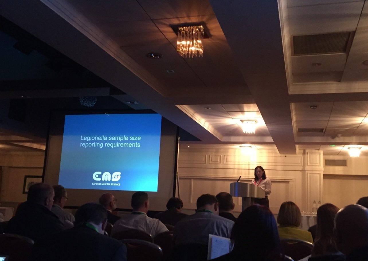
Our very own Jennifer Newton was one of the speakers at the recent LCA Open Day which was held on Thursday 2nd November 2017.

The event was a member’s oriented information day with various experts presenting in their chosen fields on Legionella and other water borne pathogens.

There was a full day’s programme which went as follows:

  • Chairman’s Welcome
  • HM Principal Specialist Inspector from HSE – Cooling Towers Intervention
  • BSi Committee Member – ISO11731:2017 Water quality – Enumeration of Legionella
  • Les Bekesi – System Design Considerations
  • Committee Member – Other Water Borne Pathogens
  • Committee Member – LCA Healthcare Standard (Compare & Contrast HSG274 and HTM04-01)
  • Jennifer Newton – Legionella sample size changes to reporting requirements
  • HM Principal Specialist Inspector from HSE – HSE enforcement rates in Healthcare
  • LCA Vice Chairman – HSG282: The control of Legionella and other infectious agents in spa-pool systems

There were around 150 participants at the event from all parts of the industry. Jennifer’s talk generated some interesting debates on how to report Legionella results, with much discussion focussing on the sample size being used.

According to ISO11731: 2017, Section 10 of this standard (Expression of results), states the following:

Report the confirmed presence (or absence) of Legionella pneumophila and the presumptive presence (or absence) of other Legionella species. Report absence as “not detectedin the volume examined. Ideally report the serogroup of all isolates of Legionella pneumophila”.

For more information, please click on Legionella sample size and reporting to see Jennifer’s blog on our technical section with highlights from her presentation.