Controlling Botulinum

Back

Controlling botulinum is a vital element of food safety that can have dire consequences if ignored.

February 24, 2022

What is Botulinum?

Botulinum, or Clostridium Botulinum, is a species of the Clostridium. This bacterium can produce toxins in low-oxygen conditions that are deadly. Low oxygen foods include, vacuum packed, canned, and foods with high oil content.

Unlike many other pathogens, labs do not routinely test for C botulinum for two reasons;

It is so deadly that testing a representative sample of food does not provide enough assurance that the entire batch is free from the pathogen.

Most labs do not have the containment levels required to propagate such a deadly bacterium.

Clostridium – Photo by CDC on Unsplash

How it is controlled

So, if routine micro testing isn’t the answer what is?

There are a few ways you can control C botulinum:

  • freezing the food, which works by inhibiting botulism rather than destroying it. This means that once the food is thawed, the botulinum can begin to take effect again, so short shelf-lives of < 10 days are required post thawing
  • high heat / temperature combinations, e.g. like those used in canning
  • having a short-shelf-life of < 10 days for product stored at temperatures > 3°C
  • maintaining a low moisture content or pH throughout the food
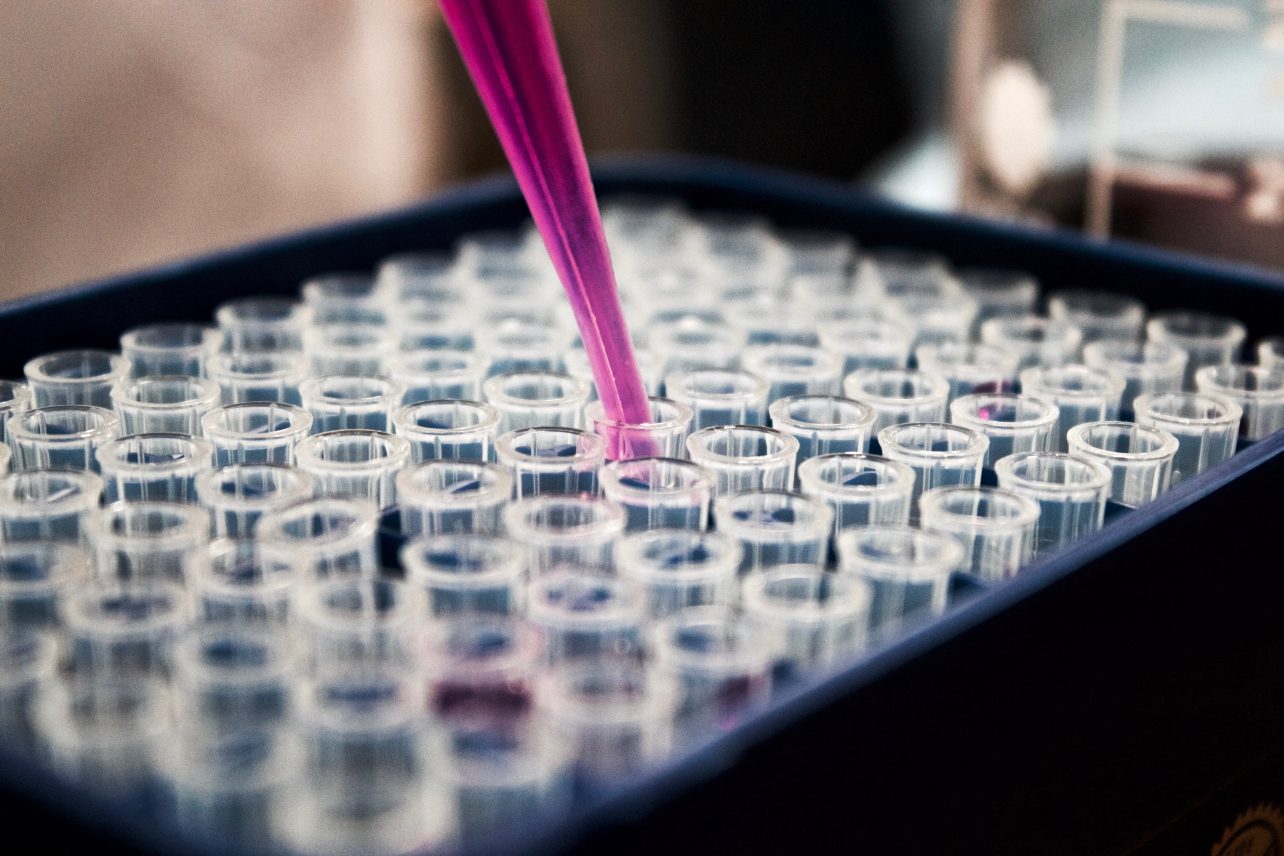
Testing

At Express Micro Science we can test your product to prove that C botulinum cannot grow under chilled conditions, so that longer shelf-lives can be achieved.

  • pH – food must be 5 or less throughout the food or throughout all components of complex foods
  • Aqueous salt – food must have 3.5% or greater of salt in the aqueous phase throughout the food or components of complex food
  • Water activity (aw) – food must have 0.97 aw or less throughout the food and throughout all components of complex foods

If your product meets any of the above requirements you are safe to extend your product’s shelf-life beyond 10 days.

A Bug’s Life: Staphylococcus Aureus

Back

Staphylococcus is a common bacteria that has many strains which can cause skin infections, food poisoning and even Toxic Shock Syndrome.

February 17, 2022

Staphylococcus, or Staph., is a type of bacteria most often found in the upper respiratory tract as well as on our skin. Most people will have some of these bacteria on their bodies – which allows for the mildly amusing anecdote that staph is found – on the Staff! It is a common skin infection and can be present in abscesses, and sinus infections (like sinusitis). There are different strains of Staph. including Staphylococcus aureus and Staphylococcus epidermidis.

At EMS, we test for Staphylococcus aureus, which can cause food poisoning by developing toxins in food and when ingested causes food poisoning. Symptoms develop within 1-6 hours and last anywhere from 30 minutes to 3 days. Food poisoning caused by Staphylococcus aureus is one of the quicker routes to a sore stomach.

Outbreaks of food poisoning caused by Staphylococcus aureus are rare, especially documented cases, due to the quick onset and recovery of victims. However, any outbreak is usually associated with products that are subject to handling during preparation or dairy products which can become contaminated from the udders of cows and sheep.

How can you prevent spreading Staph.? Wash your hands thoroughly with soap and water, especially before preparing food. If you are ill, avoid preparing food, and cover any open wounds on the hands or wrists with gloves.


The Micro Profile:

Name: Staphylococcus Aureus

Family: Staphylococcaceae

Type of ‘Bug’: Bacteria

Appearance under a microscope: grape-like clusters with round, golden-yellow colonies


History

First discovered in 1880 by Scottish scientist, Alexander Ogston, while performing a procedure. He described the bacteria as looking like “bunches of grapes”. Later, in 1884, Friedrich Julius Rosenbach found that he could differentiate the bacteria by the colour of their colonies: Staph. Aureus was named because of it’s golden colouring.


How do EMS test for Staphylococcus?

The method we use the most to test for Staph, which we are UKAS accredited to carry out is Method No 16 based on BS EN ISO:6888: Part 1: 1999 using Staphytec plus latex confirmation. If you want more information on this method, you can find it here.

The microbiology behind the bread

Back

The science in your sourdough!

April 20, 2021

By Antonio Baena Marin

As many others, this lockdown has made me my found new hobbies to do at home, so I decided to take my father in law’s advice, and I started to make my own Sourdough Bread with little hope on the result. I was shocked when I realised how the process of making bread is so similar to my job, but instead of working in the lab it takes place in the kitchen bench.

The first thing I had to do was growing my own Sourdough Starter. That took around 5 days and for that I only needed to add an equal ratio of wholemeal flour and water (Yes! As simple as that). Whole grain flour contains Wild Yeast and Lactobacilli which are the organisms needed in the fermentation of the bread. All you are looking for is to create a nice community of wild yeast and Lactobacilli that will create a symbiosis between them.

Once the starter is ready and active (Figure 1), we can start preparing our bread which is basically a mixture of sourdough started, water, salt and flour. Once the dough is mixed and kneaded, you just have to leave it and the microorganism will work their magic (also known as fermentation in the lab). That can take from 2 to 6 hours depending on the temperature of the room.

Figure 1. Sourdough Starter. Inactive Starter in the left and Active Starter in the right (red line marks the level at the start).

As microbiologist, we know that the optimum temperature for yeast to grow is 25oC, so we are aiming for that temperature. This is very difficult to achieve in cold countries like Scotland, especially in winter, but you can always place your dough in a sunny spot, next to the heater or inside your oven/microwave with the lights on (this could be considered our small incubator, like the ones we have in the lab). During this process, the dough should have risen by 30-50% (Figure 2). All is happening inside the bread is two different kinds of fermentation: Wild yeast will perform an alcoholic fermentation using the sugars; yeast will produce ethanol that will be mostly evaporated while baking the bread and CO2 that will be trapped on the gluten structure creating those nice and characteristic holes in the baked bread and causing the dough to rise. Lactobacilli digest the sugars and produce lactic acid that will lower the pH of and give that nice sour flavour to the bread.

Figure 2. Comparison of two bread doughs. Non-well fermented dough on top of image (not much rise after the mixture of ingredients) and properly fermented dough at the bottom of image (around 50% rise after mixture of ingredients).

Once the bread has doubled size, it is shaped and placed into the fridge for up to 20 hours in order to continue the process with a slow fermentation (by lowing the temperature the microorganisms will reduce their activity making the process much slower than when it takes place at around 25 oC) that is going to improve the benefits of our bread. It has been proven that slow fermentations help to preserve the bread (the low pH of the bread prevent the growth of other non-desirable microorganism and mould) avoiding the addition of additives to the bread. It also helps to reduce the gluten content since the gluten proteins are degraded, making the bread easier to digest compared to the commercial bread, where only a quick alcoholic fermentation takes place, enough for the dough to rise but without the other benefits explained above. It is also known that Lactic acid decreases the content of phytate, which inhibits iron and zinc absorption, increasing the nutritional benefits of our homemade bread.

What it is left to do is baking the bread and enjoy it.

Why are dilutions important?

Back

Check out another time lapse video explaining why dilutions are so important.

April 6, 2021

By Michael Beavitt

Accurate dilutions are one of the best used tools in microbiology for determining the number of microbes in something. No matter how many bacteria are in your initial sample, you can always dilute it down to a countable level.

In the timelapse video shown, a high concentration of E.coli was suspended in agar on the top left plate. From left to right, each plate contains a 1 in 10 dilution of that high concentration solution, then a 1 in 10 dilution of that, and so on and so forth.

Footage by Michael Beavitt

Plate one (top left) could be an example of a food sample tested in our lab with a very high bacterial load. After taking 10g of food and homogenising it with diluent, in the case of food tests, one millilitre of this solution would be added to agar, and incubated.

Each blue-green dot you see in the agar is representative of a single bacterium, which has grown into a colony. At a dilution of 1/10, there are far too many bacteria to be counted. They form a confluent lawn of growth across the entire medium. 1/100, and there’s still far too many.

The sample needs to be diluted all the way down to 1/1000000 from the original 10g suspension to allow countable colonies. Then, all it takes is some quick arithmetic to work out how many were in the 10g of food! The bottom right plate, at a dilution of 1/1000000, had 44 colonies. This means that 44000000 bacteria were present in 10g of food.

It’s very rare that anything will have this much E.coli growing on it – it was just used as an example in this case, because it’s easy to grow, and brightly coloured on the agar used. Ordinarily only a 1/10 dilution will suffice for food products! The same principles apply for other enumerations, however – an aerobic colony count will often require 1/1000 or 1/10000 for raw meat and other high risk food types.

If you’re interested, the video was shot for 24 hours, and the E.coli was incubated at 37ºC on TBX agar. Normally they’re incubated at 41.5ºC, but unfortunately my camera is not rated for those kinds of temperatures! The reason that the top 3 plates begin to crack and shrink is because all the lids were left off for ease of filming, allowing the water in the jelly to evaporate. The prolific bacterial growth also sped up the process.

Salmonella enterica Time-lapse

Back

There are many different types of Salmonella and they don’t all look the same. This is a short and sweet insight into what our microbiologists do each day in the form of a time-lapse. Enjoy!

March 2, 2021

By Michael Beavitt

Salmonella enterica subsp. enterica is one of the best-known food-poisoning culprits. While many associate it with undercooked chicken meat, in fact a large number of cases stem from the consumption of contaminated meat in general, as well as eggs and egg-containing products, and even sometimes fresh vegetables and fruit.

While a number of pathogenic varieties of Salmonella enterica exist, the most common are the Typhimurium and Enteritidis serovars. They account for the vast majority of food poisoning cases in the UK and can cause some pretty nasty symptoms.

Once they get into the body, they can trick the macrophage immune cells into “eating” them, before escaping the deadly digestive enzymes and hitching a ride to a multitude of organs and infectable sites. They can also trigger debilitating immune over-reactions, as well as producing large quantities of toxins.

Shown is a 48-hour time lapse of Salmonella enterica growing on XLD agar, which is used in our lab in the early confirmation stages of salmonella detection. One change that’s obvious is that the agar turns from orange/red to pink– this is due to the fermentation of xylose sugar and the decarboxylation of lysine (the “X” and “L” in XLD), turning the previously pH neutral surroundings alkaline. The pink colour comes from the indicator phenol red, which changes colour depending on the pH of the surrounding media. 

It’s important to note that not all Salmonella species look like this – some ferment other sugars to produce acid, turning the agar a bright yellow colour.

The other change of note is the formation of black spots in the centres of the colonies – Salmonella enterica species commonly produce H2S, a black precipitate, from sources of iron and sulphur. This allows you to quickly tell typical Salmonella species apart from other unwanted background flora that might still grow on the media.

Due to the various ways different Salmonella serovars grow on the XLD (and other selective agars used) it requires experienced microbiologists to interpret the plates as having or not having Salmonella present, and thus verifying if a food product is safe or not safe to eat.

Oysters: Dealing with Norovirus

Back

“If seven in 10 oysters have norovirus, should we still be eating them?”

February 23, 2021

By Jennifer Newton

It’s not often that I get enough time to ponder the specifics of food poisoning from molluscs. But, in the midst of feeling unwell after eating oysters, I found myself doing just that: wondering about the main contributors to food poisoning from oysters (in between trips to the bathroom, that is). A quick google search came up with “Nearly 200 ill in UK after eating oysters”, February 2020. But this is February 2021, so did I also fall ill to the same pathogen?

In the aforementioned article, the culprit was identified as the norovirus. The norovirus symptoms include:

  • Nausea.
  • Vomiting.
  • Stomach pain or cramps.
  • Watery or loose diarrhoea.
  • Feeling ill.
  • Low-grade fever.
  • Muscle pain.

Yep, I had all of them. So, looking pretty likely that I was being affected by the same pathogen that brought 200 people to their knees only a year previous. How did I get there? Well, I ate some raw oysters for Valentine’s day, and just over a 24 hours later I was feeling very poorly, which is the typical incubation period of the sickness (one to two days). And I felt unwell for just over a day, which is also in line with its classic diagnosis of lasting 1 to 3 days. The good news is; according to the CDC, people have temporary immunity from re-infection for up to 2 to 3 years.

Based on electron microscopic (EM) imagery, this three-dimensional (3D) illustration provides a graphical representation of a single norovirus virion, set against a beige background. The different colours represent different regions of the organism’s outer protein shell, or capsid.
Image by CDC

This led me to another line of questioning: I ate the oysters raw, but would I have been safe if I cooked them? Quick steaming oysters will not kill norovirus and other pathogens, according to the U.S. Centers for Disease and Prevention. To be safe, seafood must be cooked to an internal temperature of at least 145 degrees Fahrenheit (63°C). Other common sources of norovirus besides oysters include:

  • contaminated foods
  • shellfish
  • ready-to-eat foods, such as salads, ice, cookies, fruit, and sandwiches, that a worker with a norovirus infection has handled
  • any food that contains particles of the faeces or vomit of a person with norovirus

Norovirus is thought to be the most common cause of acute gastroenteritis (diarrhoea and vomiting illness) around the world. It spreads easily through food and drink and can have a big impact on people’s health. It was originally called the Norwalk virus, after the town of Norwalk, OH, where the first confirmed outbreak happened in 1972.

Can we test for norovirus?

Several methods have been developed to extract and test for total norovirus contamination (infectious and non-infectious virus particles) in foods; however, there are no internationally recognized standard methods to date. Despite improvements in our ability to extract viruses from foods, the analysis of rinses and extracts leaves much to be desired. Additionally, the method used to detect norovirus is based on PCR (Polymerase Chain Reaction) which can be costly and not normally a viable option to producers as a screening test.

“If seven in 10 oysters have norovirus, should we still be eating them?” Of course, there is a risk from eating oysters – they are harvested from the wild, after all. But the oyster itself is not the culprit, rather the water in which it is raised. All the oysters sold in the UK are purified for 42 hours, which largely nullifies any danger. But, to be completely safe, you can always cook oysters – or easier still, just zap them in a microwave, which would kill any residual traces of the virus.

So, should we still be eating oysters? Of course: they are good for you, and tasty, too!

Nutrition Facts: The Science Behind the Label

Back

All about nutritional values and how they are produced.

February 12, 2021

By Francisca Navarro Fuentes

Have you ever read the nutrition facts on your Friday nights double pepperoni frozen pizza? Did you ever wonder what those values mean, or think about how they are produced? Feed your curiosity with this post and find out about the science behind the label!

Nutrition facts are a powerful tool to help you understand the composition of a product, so that the consumer of the product is informed. Nowadays, the variety of products to choose from is endless, but knowing what is in your food can help you to pick what to buy.

By law, it is mandatory that nutrition facts are included in labels. This is controlled by Regulation (EU) No 1169/2011 [1] on the provision of food information to consumers. Therefore, nutrition declaration must include energy (KJ/Kcal), total fat and saturated fat (g), carbohydrates and sugars (g), proteins (g) and salt (g). Besides, it is optional to include values such as mono-unsaturates and polyunsaturates, polyols, starch, fibers and vitamins or minerals. The values are reported per 100g or 100mL of product, and often per portion too.

But do all products have to have a nutrition label? It may surprise you to learn that the answer is: no. There are products  exempt from this rule, such as waters, spices, salt, sweeteners, tea, food additives, gelatine, yeast and chewing gums, among others.

Most people don’t pay attention to the nutritional information and one of the reasons could be a lack of knowledge on how to interpret them. In order to help the consumer, the NHS has published a list of guidelines, summarized in Table 1.

So how are nutritional values produced?

Nutritional values are produced in analytical chemistry laboratories. A representative portion of the product arrives to the laboratory, this is then blended and homogenized into a paste (like your morning vegetable smoothy, ew!). This paste is stored in an airtight bag, ready for analysis.

Chromatography, flame photometry, spectrophotometry, high temperatures (up to 600 ºC!) and high pressures are techniques and conditions used to determine nutritional values. As you would imagine, in order to provide this information, trained chemistry analysts are required. I know, these techniques sound scary but, believe me, chemists love them! Although these machines are subject to break downs, chemists like me thrive on a broken machine’s challenge.

Now, how can you (or I), as a consumer, trust the values in the nutrition facts table? For that, a governmental body, known as United Kingdom Accreditation Service (UKAS), assesses and accredits analytical chemistry laboratories ensuring that the values obtained in laboratory A would be the same as the ones in laboratory B across the UK.

In conclusion, labels are educational as it helps to understand calories and nutrients. They regulated by Regulation (EU) No 1169/2011 and the values are generated in UKAS accredited analytical chemistry laboratories, ensuring the customer rights and accuracy of the data.

References

[1] EUR-Lex Website: https://eur-lex.europa.eu/legal-content/EN/TXT/?uri=CELEX:02011R1169-20180101 Last visited 6/2/21

[2] NHS Website: https://www.nhs.uk/live-well/eat-well/how-to-read-food-labels/#:~:text=Nutrition%20labels%20are%20often%20displayed,certain%20nutrients%2C%20such%20as%20fibre. Last visited 6/2/21